|
|
 |

|
|
|
 |
| |
-電子顕微鏡-
|
| |
|
Immunoelectron Microscopy
Methods and Protocols
Series: Methods in Molecular Biology, Vol. 657
Schwartzbach, Steven D.; Osafune, Tetsuaki (Eds.)
1st Edition., 2010, XI, 352 p. 82 illus., 41 in color., Hardcover
ISBN: 978-1-60761-782-2

Due: July 29, 2010
152,01$ |
| |
About this book
Immunoelectron microscopy is a key technique that bridges the information
gap between biochemistry, molecular biology, and ultrastructural studies
placing macromolecular functions within a cellular context. In Immunoelectron
Microscopy: Methods and Protocols, expert researchers combine the
tools of the molecular biologist with those of the microscopist. From
the molecular biology toolbox, this volume presents methods for antigen
production by protein expression in bacterial cells, methods for epitope
tagged protein expression in plant and animal cells allowing protein
localization in the absence of protein specific antibodies as well
as methods for the production of anti-peptide, monoclonal, and polyclonal
antibodies. From the microscopy toolbox, sample preparation methods
for cells, plant, and animal tissue are presented. Both cryo-methods,
which have the advantage of retaining protein antigenicity at the
expense of ultrastructural integrity, as well as chemical fixation
methods that maintain structural integrity while sacrificing protein
antigenicity have been included, with chapters examining various aspects
of immunogold labeling. Written in the highly successful Methods in
Molecular Biology? series format, chapters include introductions to
their respective topics, lists of the necessary materials and reagents,
step-by-step, readily reproducible laboratory protocols, and notes
on troubleshooting and avoiding known pitfalls. Authoritative and
essential, Immunoelectron Microscopy: Methods and Protocols seeks
to facilitate an increased understanding of structure function relationships.
Content Level " Professional/practitioner
Keywords " Fixation protocols - Macromolecular functions - Pre-
and post-embedding immunogold labeling - Structure function relationships
Related subjects " Cell Biology - Immunology |
| |
|
| |
|
| |
電子顕微鏡学 |
|
|
| |
1932年、 ドイツで電子顕微鏡(電顕)の基礎が確立された。
その後、 現在の 3段式レンズ系電顕が開発され、 その実用化が始まったのは 1950年頃で、 戦後のことである。 すなわち、
わが国は明治維新から今日に至るまで、 欧米の文化や学問を"教科書"とし、 それに追いつき、追い越せのパラダイムで支えられてきた。
そのような中にあって 「電子顕微鏡学」 は、 わが国が欧米諸国の大学と"学問のスタートライン" を同じくする、
極めて数少ない学問分野の一つである。 今日、 わが国の電顕は学問のレベル、 論文の質、 論文の発表数、 さらには日本製の電顕、
電子機器等の信頼度は海外で高い評価を受けている。 そして、わが国の電顕の生産台数は遥かに世界をリードしている。
電子顕微鏡学は、 今後、 若手研究者が欧米の諸大学と "対等な立場"に立って、 学問の発展に貢献できる、
まさに格好の分野ではないかと期待される。 |
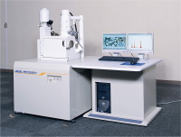
日体大・電顕室
走査型電子顕微鏡 JEOL JSM-6460LV |
|
| |
|
| |
|
| |
電子顕微鏡室 |
|
|
|
|
|
本学の電子顕微鏡室には、
透過型電子顕微鏡と付属走査型電子顕微鏡が設置されている。 電子顕微鏡(電顕)は主に研究用として大学教員、 研究員によって利用され、
数多くの研究報告が行われている。 また学生は、電顕の操作技術や基礎理論のトレーニングを受けた後、 卒業論文の作成等に使用している。
電顕室では電顕の知識、資格を有する有志によって、電子顕微鏡技術講習会や電顕の専門家を招いての勉強会が行われている。 更に、この講習会で電顕の基礎理論、電子物理、鏡体の基本操作や写真技術等の修得が認められれば、日本電子顕微鏡学会が毎年11月に実施している
「電子顕微鏡技士」 の受験資格が得られる。 現在、 本学からは田中和幸助手、 三星暢公研究員(現在、 つくば総合研究所・研究員)の
2名が難関の試験に合格し、 電顕を応用した研究に取り組んでいる。
|
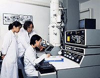
健志台キャンパス電子顕微鏡室で卒業研究に取り組む学生たち |
|
| |
|
| |
|
|
|
| |
|
第53回 日本電子顕微鏡学会学術講演会
写真コンクール 「優秀賞」 受賞作品
-ミクロンの象-
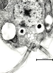
試料:単細胞緑藻
Chlamydomonas reinhardtii(クラミドモナス)
クラミドモナスは淡水や海水に棲み、光合成によってエネルギーを得ている。形態は直径約8μmの楕円体で、2本の鞭毛をもち水中を泳ぐことができる。
写真をクリックすると拡大写真がご覧いただけます。
|
電子顕微鏡の周辺機器
| ・走査型電子顕微鏡装置 JSM-6460LV |
| ・急速凍結置換固定装置 Leica EM-CPC |
| ・凍結超薄切片装置 Leica EM FCS |
| ・超薄切片装置 Leica Ultracat UCT |
| ・超薄切片装置 Porter Blum MT-1 |
| ・高画質ピクトロスタット Fuji Film 330型 |
| ・ピクトロマイティ Fuji Film A3 |
| ・自動電顕標本作成装置 A-1 |
| ・自動電顕試料作製装置 サクラ精機 HEM-480 |
| ・超薄切片装置 C.Reichert Om-U3 |
| ・コンピュタ・グラフィックス画像解析装置 |
| ・ダースト引伸機 LAB-1200 |
|
|
| |
|
|