|
|
 |
 |
|
|
 |
| �@ |
-���ۊw��̕\��������-
|
| �@ |
�ʐ^���N���b�N����Ɗg��ʐ^���������������܂��B
|
| �@ |
�@ |
| �@ |
 |
| �@ |
Immunoelectron Microscopy
Methods and Protocols
Series: Methods in Molecular Biology, Vol. 657
Schwartzbach, Steven D.; Osafune, Tetsuaki (Eds.)
1st Edition., 2010, XI, 352 p. 82 illus., 41 in color., Hardcover
ISBN: 978-1-60761-782-2

Due: July 29, 2010
152,01$
�@
About this book
Immunoelectron microscopy is a key technique that bridges the information
gap between biochemistry, molecular biology, and ultrastructural
studies placing macromolecular functions within a cellular context.
In Immunoelectron Microscopy: Methods and Protocols, expert researchers
combine the tools of the molecular biologist with those of the microscopist.
From the molecular biology toolbox, this volume presents methods
for antigen production by protein expression in bacterial cells,
methods for epitope tagged protein expression in plant and animal
cells allowing protein localization in the absence of protein specific
antibodies as well as methods for the production of anti-peptide,
monoclonal, and polyclonal antibodies. From the microscopy toolbox,
sample preparation methods for cells, plant, and animal tissue are
presented. Both cryo-methods, which have the advantage of retaining
protein antigenicity at the expense of ultrastructural integrity,
as well as chemical fixation methods that maintain structural integrity
while sacrificing protein antigenicity have been included, with
chapters examining various aspects of immunogold labeling. Written
in the highly successful Methods in Molecular Biology? series format,
chapters include introductions to their respective topics, lists
of the necessary materials and reagents, step-by-step, readily reproducible
laboratory protocols, and notes on troubleshooting and avoiding
known pitfalls. Authoritative and essential, Immunoelectron Microscopy:
Methods and Protocols seeks to facilitate an increased understanding
of structure function relationships.
Content Level " Professional/practitioner
Keywords " Fixation protocols - Macromolecular functions -
Pre- and post-embedding immunogold labeling - Structure function
relationships
Related subjects " Cell Biology - Immunology
|
| �@ |
|
| �@ |
���[�O���i�� "����t�Α�"�̔���
|
| �@ |
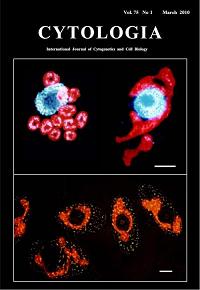
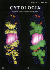
Formation of "giant chloroplast" during the cell cycle
of Euglena gracilis Z in synchronized culture
Osafune,T,,Tanaka,K. and Ehara,T.
Cytorogia, Vol.74,1,(2010)
In the cell cycle of Euglena gracilis Z in synchronized cultures,
chloroplasts temporarily conjoin to form a single giant structure
at the 14th hour after the onset of the light period, called "giant
chloroplast" (upper in right and bottom), which surrounds the
nucleus making connections or close contacts at several sites. The
upper left shows the 10th hour after the onset of the light period.
The cell contained 11 chloroplasts and chloroplasts were round or
oval in shapes and dispersed in the cytoplasm. Chloroplast nucleoids
in the "giant chloroplast", observed under a fluorescence
microscope after staining with DAPI, the DNA fluorochrome, become
stringy and some tips of the string appeared to come into close
proximity to the site of connection with the nucleus (upper in right).
Bar (upper, bottom: 5mm).
|
| �@ |
|
| �@ |
�����|�{���[�O���i��"����t�Α�"�̌`�� |
| �@ |
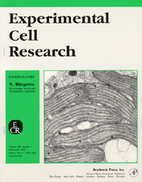
Ehara,T.,Osafune,T.,Hase, E.:
Interactions between the nucleus and cytoplasmic organelles during
the cell cycle of Euglena gracilis in synchronous cultures.
Exp. Cell Res.190(1)104-112, (1990)
�@�@��ɁAOsafune��͒P�זE���חΑ��N���~�h���i�X�̓����|�{�W�c�ɂ�����~�g�R���h���A�̌`�Ԃ�ǐՂ��A�����̏��^�~�g�R���h���A�����ݗZ���ɂ���ċ���~�g�R���h���A�`������A
�����Ɍċz�������ꎞ�I�ɒቺ���錻�ۂ��݂��������B �������́A���̂悤�Ȍ��ۂ͗t�Α̂ł��N�肤����̂Ɛ������A�����̗t�Α̂�L����P�זE�ږё����[�O���i�Œ��������B
���̌��ʁA ���[�O���i��cell cycle���̏����ƍזE�����O�ƂɁA�ꎞ�I��"����t�Α�"���o�������Ƃ��݂��������B
�����āA �t�Α̂̃_�C�i�~�b�N�ȓ��Ԃ��ŏ���cell cycle���Ɉʒu�t�����B
|
| �@ |
|
| �@ |
���[�O���i��LHCII�@�^���p�N�����q�̓S���W�̂��o�R���� |
| �@ |
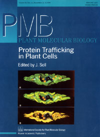 �@ �@
Schwartzbach,S.D., Osafune, T., Löffelhardt, W.:
Protein import into cyanelles and complex chloroplasts.
Plant Molecular Biology. 382(2)263-274 (1998)
�@�@�����w�nII�Ɍ����W�߂�W�����N�����t�B��a/b�����^���p�N��������(LHCII)�͍זE�j�ɃR�[�h����čזE���ō�������A
���炩�̕��@�ɂ���ėt�Α̂ɉ^�ꂽ��A 2�i�K�̃v���Z�b�V���O���ă`���R�C�h���ɑg���܂�邱�Ƃ��m���Ă���B�������̓��[�O���i���|�{���A
LHCII�@�^���p�N�����q�̍זE���̓��Ԃ�Ɖu�d���@�ƃR���s���[�^�E�O���t�B�b�N�X�ƂŒǐՂ��A LHCII�^���p�N�����q���S���W�̂��o�R����,
�t�Α̃`���R�C�h���ɗA������錻�ۂ��ŏ��ɂ݂������� (Osafune,et al.:Exp.Cell Res.193:320-330,1991)�B
���̂悤�ȐV�����͒N���\�z���Ȃ����������ŁA����̐V���Ȃ錤�����W�J������̂Ǝv����B�@
|
| �@ |
|
| �@ |
���[�O���i�̃v���X�`�h�ƃv���������̂�3�����č\�z |
| �@ |
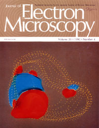 �@ �@
Osafune,T.,Ehara,T.,Sumida,S.,Hase,E., Schiff,J.A.:
Light-independent processes in the formation of thylakoids and pyrenoids
in proplastids of dark-grown cells of Euglena gracilis.
J. Electron Microscopy. 39(4)245-253, (1990)
�@�@��ɁA Euglena gracilis Z�͓K�ȑO�|�{������I�ׂA
�Ï��Ńv���v���X�`�h�\���̔��B���N���邱�Ƃ��݂��������B�@ �{�_���͘A�������ؕЖ@-�d�q�������A �R���s���[�^�E�O���t�B�b�N�X�摜��͑��u��p���āA���[�O���i�̗t�Α̌`���̏����Éߒ��ɂ�����v���X�`�h�̔��\���̕ω����o���I�ɒǐՂ������̂ł���B�@�ʐ^��144���Ԍ�̃v���v���X�`�h�\���ł���B�@�]���A
�s���m�C�h�͌��̗U���ɂ��`�������ƍl�����Ă���(Klein)�B ����A �������̃O���[�v�͊T�����A�ďo�����t�Α̌`�������Éߒ��̊ώ@�\�Ȏ����n����щ摜��͑��u��p���āA�s���m�C�h���Ï��ł��`������邱�Ƃ𖾂炩�ɂ����B�@
|
| �@ |
|
| �@ |
LHCII�@�^���p�N�����q�̍זE���R�������z |
| �@ |
Osafune,T.:
A three-dimensional computer generated model of synchronously dividing
Euglena gracilis Z cell at light/dark cycle constructed based
on serial sections.
Cytologia. 67,(1)1-2,
(2000)
�@�@�����|�{����Euglena gracilis Z��Ɖu�d���@�ƃR���s���[�^�E�O���t�B�b�N�X�摜��͂ɂ��A �זE��LHCII�@�^���p�N�����q��
3�����č\�z�������̂ł���B ���F�Ɏ������h�b�g��LHCII�^���p�N�����q�ŁA�זE���̔z�u��3�����I�ɕ\������Ă���B���Ȃ킿�A LHCII�͗t�Α̂ƑS�ẴS���W�̂ɋǍ݂��Ă��邱�Ƃ�����B
3�����I�ɍ\�z���ꂽ���̓f�B�X�v���C��ʏ�ŔC�ӂ̊p�x�Ŋ��f���A �f�ʑ��̊ώ@��̊g��A�k���A �ړ����]�����邱�Ƃ��\�ł���B
���F�h�b�g��LHCII�^���p�N�����q�B �s���N�͍זE�j�A �ΐF�͗t�ΆA �ԐF�̓S���W�́A �F�̃h�b�g�͍זE�����������Ă���B
|
| �@ |
|
| �@ |
���[�O���i��RuBisCO�y�f�̍זE�� 3�������z |
| �@ |
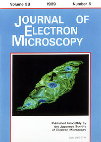 �@ �@
Osafune,T.,Sumida,S.,Ehara,T.,Hase,E.:
Three-dimensional distribution of ribulose-1,5-bisphosphate carboxylase
oxygenase in chloroplasts of actively photosynthesizing cell of Euglena
gracilis.
J. Electron Microscopy,38(5)399-402, (1989)
�@�@Euglena gracilis Z�זE��ribulose-1,5-bisphosphate
carboxylase oxygenase(RuBisCO)��A�������ؕЖ@�A�Ɖu�d���@����уR���s���[�^�E�O���t�B�N�X�摜��͂ɂ���āA�זE����
3�����z�𖾂炩�ɂ����B���̂悤�ȕ��q�זE�`�Ԋw�I������@�ŁA�������Y�_�Œ蔽���̎�v�y�f�ł���RuBisCO�̖� 80%���s���m�C�h�ɋǍ݂��邱�Ƃ��m�肵���B
���Ȃ킿�A ����܂ł�RuBisCO�ɂ��Ă̐����܂����߂ɁA�����������Ē�ʓI�ȉ�^�����ŏ��̘_���ł���B�h�b�g��RuBisCO�A�ΐF�͗t�Α̂������Ă���B
|
|